

Anatomy Of Foot Ankle Anatomy Of The Foot Amp Ankle Total Ankle Replacement Ankle anatomy
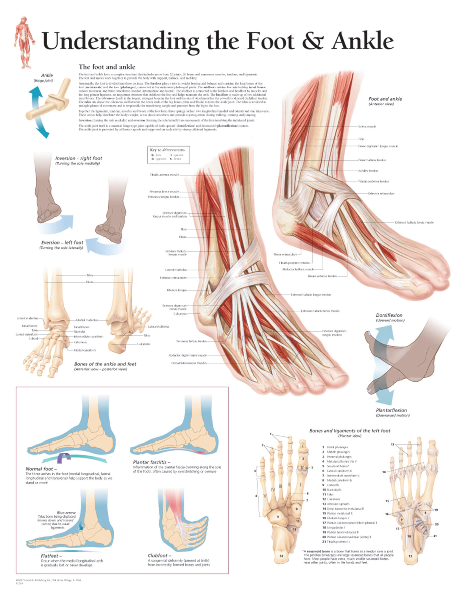
It is made up of three joints: upper ankle joint (tibiotarsal), talocalcaneonavicular, and subtalar joints. The last two together are called the lower ankle joint. The upper ankle joint is formed by the inferior surfaces of tibia and fibula, and the superior surface of talus.
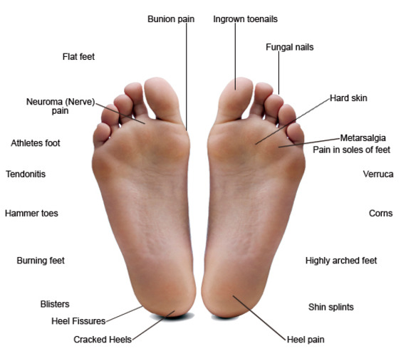
Common Foot Problems — Hawaii Podiatry
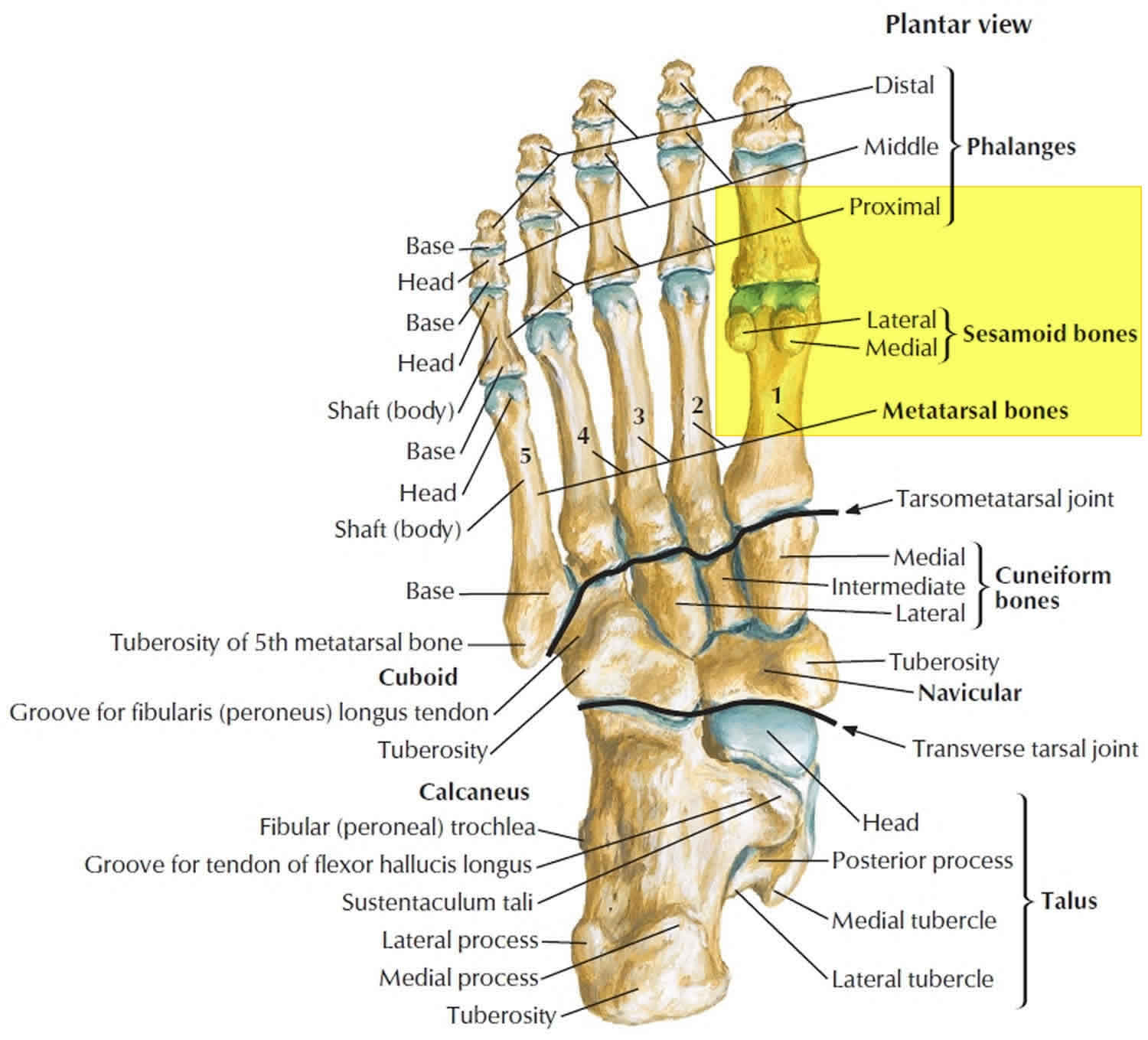
The anatomy of the foot The foot contains a lot of moving parts - 26 bones, 33 joints and over 100 ligaments. The foot is divided into three sections - the forefoot, the midfoot and the hindfoot. The forefoot This consists of five long bones (metatarsal bones) and five shorter bones that form the base of the toes (phalanges).
Bones of the human foot diagram 1142236 Vector Art at Vecteezy
It consists of 28 bones, which can be divided functionally into three groups, referred to as the tarsus, metatarsus and phalanges. The foot is not only complicated in terms of the number and structure of bones, but also in terms of its joints.
The 19 Muscles Of The Foot / The muscles of the foot Stock Image F001/4573 / The
In most two-footed and many four-footed animals, the foot consists of all structures below the ankle joint: heel, arch, digits, and contained bones such as tarsals, metatarsals, and phalanges; in mammals that walk on their toes and in hoofed mammals, it includes the terminal parts of one or more digits. The parts of a dog's hind foot and forefoot.

Foot & Ankle Bones
These bones are arranged in two rows, proximal and distal. The bones in the proximal row form the hindfoot, while those in the distal row from the midfoot. Hindfoot. Talus. Calcaneus. The talus connects the foot to the rest of the leg and body through articulations with the tibia and fibula, the two long bones in the lower leg. Midfoot. Navicular.

Chart of FOOT Dorsal view with parts name Vector image Stock Vector Image & Art Alamy
Parts of the foot. The foot is divided into three parts, structurally speaking; the forefoot, midfoot and hindfoot. Forefoot. The forefoot contains your toes and their metatarsals. Each of your toes is made of several small bones. The big toe has two bones, the proximal and distal. It also has one joint known as the interphalangeal joint.

Foot Anatomy 101 A Quick Lesson From a New Hampshire Podiatrist Nagy Footcare
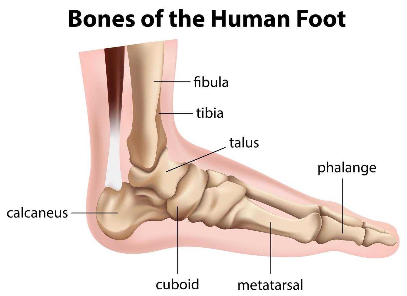
Foot Bone Anatomy tibia, fibula tarsus (7): talus, calcaneus, cuneiformes (3), cuboid, and navicular metatarsus (5): first, second, third, fourth, and fifth metatarsal bone phalanges (14) There can be many sesamoid bones near the metatarsophalangeal joints, although they are only regularly present in the distal portion of the first metatarsal bone.

Foot & Ankle Bones
The bones of the foot are organized into rows named tarsal bones, metatarsal bones, and phalanges. These make up the toes and broad section of the feet. The other bones of the foot that create.
Understanding the Foot & Ankle Scientific Publishing
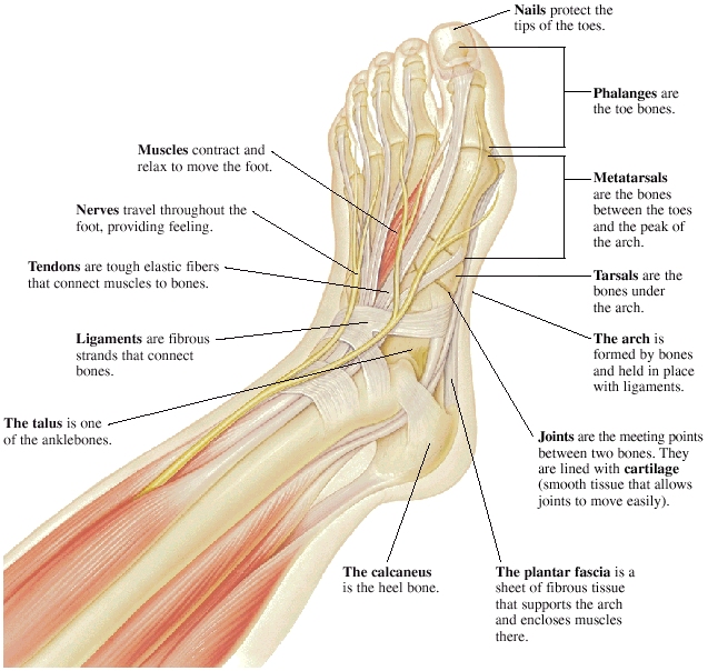
33 joints more than 100 muscles, tendons, and ligaments Bones of the foot The bones in the foot make up nearly 25% of the total bones in the body, and they help the foot withstand weight..
Turf toe causes, signs, symptoms, recovery, diagnosis & turf toe treatment
Bones of foot. The 26 bones of the foot consist of eight distinct types, including the tarsals, metatarsals, phalanges, cuneiforms, talus, navicular, and cuboid bones. The skeletal structure of.

Foot pain looking up the chain
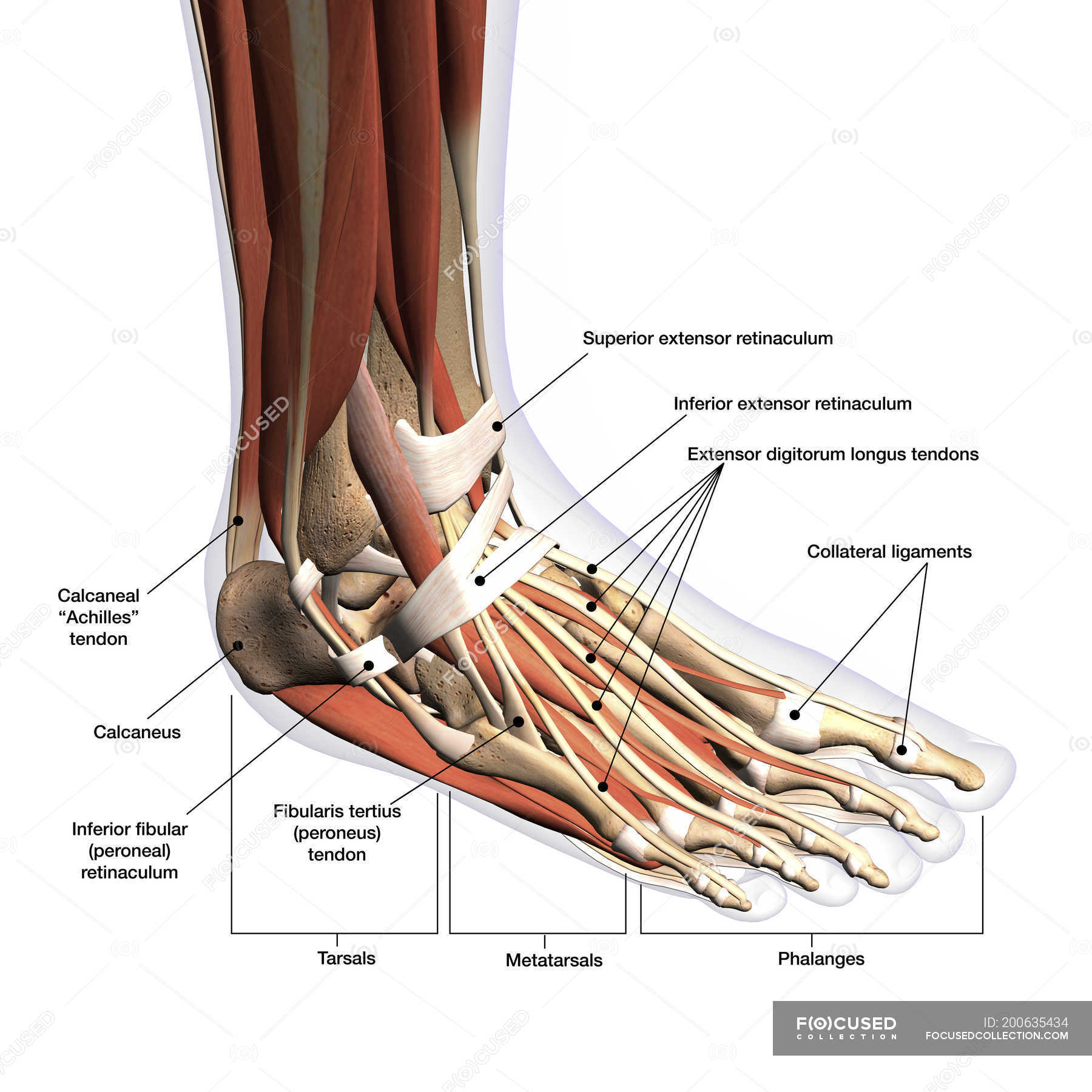
LABELED DIAGRAMS. Figure 1. Sections and Bones of the Foot A. Lateral (Left) B. Anterior (Right) Figure 2. Compartments of the Foot A. Cut Section through Mid-Foot. Figure 3. First Layer of the Foot A. Plantar View of Right Foot. Figure 4. Second Layer of the Foot A. Plantar View of Right Foot.

anatomy of the foot Ballet News Straight from the stage bringing you ballet insights
Foot Anatomy . There are many parts of the foot and all have important jobs. Each foot has 26 bones, over 30 joints, and more than 100 muscles, ligaments, and tendons. These structures work together to carry out two main functions:
Advantage Orthopedic and Sports Medicine Clinic Gresham, OR Health Library
The midfoot is a pyramid-like collection of bones that form the arches of the feet. These include the three cuneiform bones, the cuboid bone, and the navicular bone. The hind foot forms the heel and ankle. The talus bone supports the leg bones (tibia and fibula), forming the ankle. The calcaneus (heel bone) is the largest bone in the foot.

Image result for skull sketch anatomy underside Foot anatomy, Human anatomy picture, Anatomy
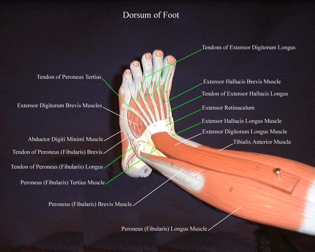
Fig 1 - The dorsal layer of foot muscles. Plantar Aspect There are ten intrinsic muscles located in the plantar aspect (sole) of the foot. They act collectively to stabilise the arches of the foot and individually to control movement of the digits. They are innervated by the medial or lateral plantar nerves - which are branches of the tibial nerve.
Anatomy of human foot with labels on white background — ankle, leg Stock Photo 200635434
The foot is one of the most complex parts of the body. It consists of 28 bones connected by many joints, muscles, tendons, and ligaments. The foot is prone to many types of injuries. Foot pain and problems can cause pain and inflammation, limiting movement. Muscles contract and relax to move the foot.

Anatomy of the Foot and Ankle OrthoPaedia
It is made up of over 100 moving parts - bones, muscles, tendons, and ligaments designed to allow the foot to balance the body's weight on just two legs and support such diverse actions as running, jumping, climbing, and walking. Because they are so complicated, human feet can be especially prone to injury.